
Function of stimulus material
Controlled stimulus material is needed when visual judgements are studied systematically.
The material basis goes back to 3D acquisitions from 2005 onward. It links morphological prior work on faces and age with questions of visual judgement, observer variability, and confidence.
Systematic control or variation of age, view, image information, and task makes it possible to distinguish more specifically between potential contributions of the face, the view, the image conditions, and the observer.
How does a visual judgement change when the same person is represented differently?
In faces, view, projection, and image conditions are not secondary. They affect which cues are visible, which are obscured, and what observers can infer from them.
Stimulus pool and age range
The stimulus pool comprises 3D face data and standardised image data from 208 people (105 women, 103 men) aged 9 to 85 years. The data allow controlled views, derived images, age-estimation tasks, comparison tasks, and analyses of visible cues.
The point is not to demonstrate scanning technology. The material provides a basis for relating stimulus variation, observer judgement, and uncertainty in a systematic way.

What can be studied
Age and visible cues
Which structures are used for age estimates, and why do estimates vary more for some people than for others?
View and projection
How do frontal, lateral, or derived views change the available information and the confidence of judgements?
Observer judgements
How consistent are judgements across observers, across repeated presentations, and in relation to subjective confidence?

3D acquisition as a basis
3D acquisition separates geometric shape, surface texture, and derived 2D views. This makes differences between facial form, image representation, and observer judgement easier to examine directly.
Technical details on acquisition
The data were acquired with a non-contact 3D laser scanner based on laser triangulation. The scanner does not interpret features; it records a dense point cloud of the object surface. These points were subsequently combined with photographic RGB information.
Each scan captured 640 × 480 measurement values, corresponding to 307,200 measurement points. The original technical documentation reports a measurement accuracy of ±0.010 mm at one sigma and a geometric z-resolution of 0.008 mm.
The scan itself took 2.5 seconds per position. For each head, three positions were acquired: lateral left, frontal, and lateral right. Focus distance was 1060 mm for the frontal scan and 920 mm for the lateral scans.

Documentation of the 3D workflow
Workflow figures
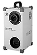


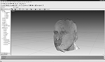
Prior work on visible ageing cues

Show additional views and legend
The prior work included frontal, lateral, and oblique isometric views. This material makes it possible to examine to what extent judgements of visible ageing cues depend on view, contour, skin relief, and soft-tissue form.

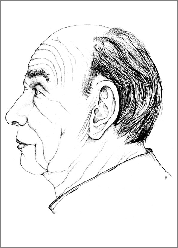

Lineae frontales transversae — horizontal forehead lines
Lineae glabellares verticales — vertical glabellar lines
Sulcus orbitalis superior — superior orbital furrow
Sulcus nasalis transversus — nasal root furrow
Sulcus anonymus superior — upper lid fold transition furrow
Sulcus orbito-palpebralis superior — upper eyelid furrow
Lineae canthi lateralis — lateral canthus lines
Sulcus orbito-palpebralis inferior — lower eyelid furrow
Sulcus anonymus inferior — canthal furrow
Lineae orbitales laterales — temporal orbital lines
Plica naso malaris — naso-malar fold
Sulcus orbitalis inferior — inferior orbital furrow
Sulcus oculo-malaris — oculo-malar furrow
Sulcus alaris superior — superior alar furrow
Sulcus alaris inferior — inferior alar furrow
Sulcus nasolabialis — nasolabial furrow
Sulcus nasooralis — philtral / naso-oral groove
Fovea buccalis — buccal dimple
Lineae supralabiales — supralabial lines
Sulcus angularis — mouth-angle furrow
Sulcus sublabialis — sublabial furrow
Sulcus mentolabialis — mentolabial furrow
Fovea mentalis — mental dimple
Lineae cervicales — horizontal neck lines
The legend names the labelled wrinkle, furrow, and line structures; it does not constitute a current scoring system.
Original format, export, and further processing
After acquisition, geometric surface data and photographic texture data were combined. The central point is that controlled views and different forms of representation can be derived from the same base material.
Show technical file formats
The original single scans are stored in the Konica Minolta/CDM format. Documented system output or export formats include STL, DXF, OBJ, ASCII points, and VRML; meshed 3D models were stored as VRML.
- Konica Minolta / CDM (original single scans)
- STL (*.stl)
- DXF (*.dxf)
- Wavefront OBJ (*.obj)
- ASCII points
- VRML (*.wrl; processed meshed models)
Protection of the material
The stimulus material is described only in general form. Person-related image data, individual case data, and raw data are not publicly displayed.